Cittasanto
  • Home
    • About Cittasanto
  • Notes
  • Reflections
  • Meditation
  • Translations
  • Acariyā
    • Quotes
    • Videos
    • Writings >
      • Writings Posts
  • Art
  • Calendar
  • Reviews
  • Lexicon
  • Links

Bone Marrow

11/30/2012

0 Comments

 
Picture
Description
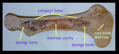
Bone marrow is the flexible tissue found in the interior of bones.

At birth, all bone marrow is red. With age, more and more of it is converted to the yellow type; only around half of adult bone marrow is red. Red marrow is found mainly in the flat bones, such as the pelvis, sternum, cranium, ribs, vertebrae and scapulae, and in the cancellous ("spongy") material at the epiphyseal ends of long bones such as the femur and humerus. Yellow marrow is found in the medullary cavity, the hollow interior of the middle portion of long bones. In cases of severe blood loss, the body can convert yellow marrow back to red marrow to increase blood cell production.

On average, bone marrow constitutes 4% of the total body mass of humans; in an adult weighing 65 kilograms (140 lb), bone marrow accounts for approximately 2.6 kilograms (5.7 lb). The hematopoietic compartment of bone marrow produces approximately 500 billion blood cells per day, which use the bone marrow vasculature as a conduit to the body's systemic circulation. Bone marrow is also a key component of the lymphatic system, producing the lymphocytes that support the body's immune system.

Function
Red blood cells are produced in the heads of long bones, in a process known as hematopoesis. The red bone marrow is a key element of the lymphatic system, being one of the primary lymphoid organs that generate lymphocytes from immature hematopoietic progenitor cells. Furthermore, bone marrow performs a valve-like function to prevent the backflow of lymphatic fluid in the lymphatic system.
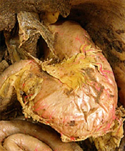
Colour
Yellow/Red.

Shape
Soft tissues like Jell-O.

Location
Marrow is found mainly in the flat bones such as the hip, breast, skull, ribs, vertebrae, and shoulder blades, as well in long bones such as the femur and tibia.

0 Comments

Spleen

11/30/2012

0 Comments

 
Picture
Description
The spleen, in healthy adult humans, is approximately 11 centimetres (4.3 in) in length. It usually weighs between 150 grams (5.3 oz) and 200 grams (7.1 oz).

The spleen is 1" by 3" by 5", weighs approximately 7 oz, and lies between the 9th and 11th ribs on the left hand side.

Function
  • Mechanical filtration of red blood cells. In mice: Reserve of monocytes.


  • Production of opsonins, properdin, and tuftsin.
  • Creation of red blood cells. While the bone marrow is the primary site of hematopoiesis in the adult, the spleen has important hematopoietic functions up until the fifth month of gestation. After birth, erythropoietic functions cease, except in some hematologic disorders. As a major lymphoid organ and a central player in the reticuloendothelial system, the spleen retains the ability to produce lymphocytes and, as such, remains an hematopoietic organ.
  • Storage of red blood cells, lymphocytes and other formed elements. In horses, roughly 30% of the red blood cells are stored there. The red blood cells can be released when needed. In humans, up to a cup (236.5 ml) of red blood cells can be held in the spleen and released in cases of hypovolemia. It can store platelets in case of an emergency. Up to a quarter of lymphocytes can be stored in the spleen at any one time.

Colour
Dark red.

Shape
An elongated sponge like tissue organ similar to a medium water balloon.

Location
In the upper left quadrant of the abdomen.

0 Comments

Heart

11/24/2012

0 Comments

 
Picture
Description
A hollow, muscular, contractile 10-ounce organ, the centre of the circulatory system. The heart pumps over 300 quarts of blood an hour. The heart beats around 100,000 times every day and some 37,000,000 times a year. The human heart creates enough pressure to squirt blood 30 feet.

Function
It provides propulsion to circulate blood throughout the vascular system.

Colour
Red.

Shape
Like a Lotus bud.

Location
Found between the lungs inside the body 2/3 to the left of middle.

0 Comments

Liver

11/24/2012

0 Comments

 
Picture
Description
Largest internal glandular organ in the body, weighing approximately 3– 3.5 pounds.

Function
The liver is first to receive blood from the intestines where the blood has absorbed the final products of digestion and decomposition. The liver removes toxic products and purifies digestion and assimilation. It incorporates amino acids into energy production. It synthesizes cholesterol as well as lipoproteins for the transport of fat to other body tissues; it converts fatty acids so they may be used as energy sources. The liver stores and releases sugar. The liver is the storage place for copper, iron, vitamin B12 and fat-soluble vitamins A, D, E, and K. The liver excretes bile.

Colour
Reddish brown.

Shape
Triangular. Convex on it’s upper margin, concave in it’s lower margin and about the size of a Rugby ball.

Location
In the upper right quadrant of the abdominal cavity resting just below the diaphragm. The liver lies to the right of the stomach and overlies the gallbladder.

0 Comments

Diaphragm

11/24/2012

0 Comments

 
Picture
Description
The thin layer of muscle that separates the chest cavity containing the lungs and heart from the abdominal cavity containing the intestines and digestive organs. It extends across the bottom of the rib cage.

Function
The diaphragm functions in breathing. During inhalation, the diaphragm contracts and moves in the inferior direction, thus enlarging the volume of the thoracic cavity (the external intercostal muscles also participate in this enlargement). This reduces intra-thoracic pressure: In other words, enlarging the cavity creates suction that draws air into the lungs.

Colour
White/Red.

Shape
Dome shaped.

Location
Base of the rib-cage.

Delimitation
The diaphragm is a dome-shaped musculofibrous septum that separates the thoracic from the abdominal cavity, its convex upper surface forming the floor of the former, and its concave under surface forming the roof of the latter. Its peripheral part consists of muscular fibers that take origin from the circumference of the inferior thoracic aperture and converge to be inserted into a central tendon.


0 Comments

Kidneys

11/23/2012

0 Comments

 
Picture
Description

There are two kidneys. Each is 4-6 ounces, 4.5 inches long, 2-3 inches broad, and 1 inch thick. About 400 gallons of blood flow through the kidneys each day.

Function
The kidneys serve several essential regulatory roles. They are essential in the urinary system and also serve homeostatic functions such as the regulation of electrolytes, maintenance of acid–base balance, and regulation of blood pressure (via maintaining salt and water balance). They serve the body as a natural filter of the blood, and remove wastes which are diverted to the urinary bladder. In producing urine, the kidneys excrete wastes such as urea and ammonium, and they are also responsible for the reabsorption of water, glucose, and amino acids. The kidneys also produce hormones including calcitriol, erythropoietin, and the enzyme renin.

Colour
Purplish and brown in Colour.

Shape
Like large kidney beans.

Location
The kidneys are located in the abdominal cavity, more specifically in the paravertebral gutter and lie in a retroperitoneal position at a slightly oblique angle. There are two, one on each side of the spine. The asymmetry within the abdominal cavity caused by the liver typically results in the right kidney being slightly lower than the left, and left kidney being located slightly more medial than the right.

0 Comments

Lungs

11/23/2012

0 Comments

 
Picture
Description
Two cone shaped spongy organs of respiration. The right lung consists of three lobes while the left lung is slightly smaller consisting of only two lobes (the left lung has a "cardiac notch" allowing space for the heart within the chest).

The lungs contain approximately 2,400 kilometres (1,500mi) of airways and 300 to 500 million alveoli, having a total surface area of about 70 square metres (750 sq ft) (8,4 x 8,4m) in adults.

Each lung weighs 1.1 kilograms (2.4lb), therefore making the entire organ about 2.3 kilograms (5.1lb).

Function
The primary purpose is to bring air and blood into intimate contact so that oxygen can be added to the blood and carbon dioxide can be removed. This is achieved by two pumping systems, one moving gas and the other liquid. During a 24-hour period, the average human will breathe 23,040 times. Humans breathe 20 times per minute, more than 10 million times per year and about 700 million times in a lifetime.

The lungs convert angiotensin I to angiotensin II. In addition, they remove several blood-borne substances, e.g. PGE1, PGE2, PGF2α, leukotrienes, serotonin, bradykinin.

Colour
Dark red.

Shape
Like two long melons cut in half. The internal surface area is similar to a honeycomb and approximately the same size as a tennis court.

Location
In the chest.

0 Comments

Large Intestine

11/17/2012

0 Comments

 
Picture
Description
The large intestine (or large bowel) is the last part of the digestive system.

The large intestine consists of the cecum, colon, rectum and anal canal. It starts in the right iliac region of the pelvis, just at or below the right waist, where it is joined to the bottom end of the small intestine. From here it continues up the abdomen, then across the width of the abdominal cavity, and then it turns down, continuing to its endpoint at the anus.

The large intestine is about 4.9 feet (1.5 m) long, which is about one-fifth of the whole length of the intestinal canal.

Function
The large intestine takes about 16 hours to finish the digestion of the food. It removes water and any remaining absorbable nutrients from the food before sending the indigestible matter to the liver. The colon absorbs vitamins which are created by the colonic bacteria - such as vitamin K (especially important as the daily ingestion of vitamin K is not normally enough to maintain adequate blood coagulation), vitamin B12, thiamine and riboflavin. It also compacts faeces, and stores faecal matter in the rectum until it can be discharged via the anus in defecation.

Colour
White.

Shape
Upside down U Shaped, like a thick snake.

Location
Within the abdomen between the small intestines and anus.

Parts of the large intestine are:
  • Cecum – the first part of the large intestine
  • Taeniae coli – three bands of smooth muscle
  • Haustra – bulges caused by contraction of taeniae coli
  • Epiploic appendages – small fat accumulations on the viscera
Locations along the colon are:
  • The ascending colon
  • The right colic flexure (hepatic)
  • The transverse colon
  • The transverse mesocolon
  • The left colic flexure (splenic)
  • The descending colon
  • The sigmoid colon – the v-shaped region of the large intestine


0 Comments

Small Intestine

11/17/2012

0 Comments

 
Picture
Description
The average length of the small intestine in an adult human male is 6.9 m (22 feet 6 inches), and in the adult female 7.1 m (23 feet 4 inches). It can vary greatly, from as short as 4.6 m (15 feet) to as long as 9.8 m (32 feet). It is approximately 2.5–3cm in diameter.

The small intestine is divided into three structural parts: the Duodenum; Jejunum; and Ileum.

Function
The small intestine is where most chemical digestion takes place.The three major classes of nutrients that undergo digestion are proteins, lipids (fats) and carbohydrates. Digested food then able to pass into the blood vessels in the wall of the intestine through the process of diffusion. The small intestine is the site where most of the nutrients from ingested food are absorbed.

Absorption of the majority of nutrients takes place in the jejunum, with the following notable exceptions:
  • Iron is absorbed in the duodenum.
  • Vitamin B12 and bile salts are absorbed in the terminal ileum.
  • Water and lipids are absorbed by passive diffusion throughout the small intestine.
  • Sodium bicarbonate is absorbed by active transport and glucose and amino acid co-transport.
  • Fructose is absorbed by facilitated diffusion.

Colour
White.

Shape
Like a very long coiled snake.

Location
From the duodenum to the large intestine within the abdomen.

0 Comments

Stomach

11/17/2012

1 Comment

 
Picture
Description
The stomach is a muscular, hollow, dilated part of the digestion system which functions as an important organ of the digestive tract in some animals, including vertebrates, echinoderms, insects (mid-gut), and molluscs. It is involved in the second phase of digestion, following mastication (chewing).

Function
Bolus (masticated food) enters the stomach through the oesophagus via the oesophageal sphincter. The stomach releases proteases (protein-digesting enzymes such as pepsin) and hydrochloric acid, which kills or inhibits bacteria and provides the acidic pH of two for the proteases to work. Food is churned by the stomach through muscular contractions of the wall called peristalsis – reducing the volume of the fundus, before looping around the fundus and the body of stomach as the boluses are converted into chyme (partially digested food). Chyme slowly passes through the pyloric sphincter and into the duodenum of the small intestine, where the extraction of nutrients begins. Depending on the quantity and contents of the meal, the stomach will digest the food into chyme anywhere between forty minutes and a few hours. The human stomach can hold approximately 1.5 litres of food.

Colour
Clear with whatever the colour of the food contents.

Shape
A "J" shaped pouch.

Location
The stomach is located in the upper left of the abdomen between the esophagus and the small intestine sitting just below the diaphragm.

1 Comment
<<Previous
Forward>>

    About

    This Document has been put together using information gathered from Wikipedias Patikulamanasikara page links found in the section “Objects of contemplation” or the website “The 32 Parts of the Body”.

    Photographs obtained from Google, Yahoo, or file images from research by myself & others in the past.

    As this is a work-in-progress information from different sources will be added as time allows, or becomes available, so please comment upon these posts.

    Archives

    December 2012
    November 2012

    Categories

    All

    RSS Feed

Powered by Create your own unique website with customizable templates.